MRIMRI means Magnetic Resonance Imaging but it can also mean the exam, the image, or the imager. Further information on MRI and its technique More, a revolutionary technique!
MRIMRI means Magnetic Resonance Imaging but it can also mean the exam, the image, or the imager. Further information on MRI and its technique More is a fairly new medical imaging technique as it has been used in hospitals and others health care facilities since the 1980s. It reveals images, with a high degree of accuracy, of most of the organs: brain, heart, spine, joints, soft tissues, etc.
From NMR to MRIMRI means Magnetic Resonance Imaging but it can also mean the exam, the image, or the imager. Further information on MRI and its technique More: physical principle
MRIMRI means Magnetic Resonance Imaging but it can also mean the exam, the image, or the imager. Further information on MRI and its technique More is based on a physical phenomenon named Nuclear Magnetic Resonance (NMR).
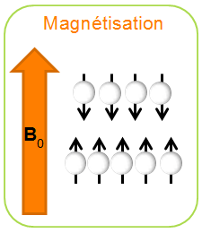
| The “Nuclear” part of the acronym NMR come from the fact that we focus on specific properties of the NUCLEUS of atoms (“nucleus” comes itself from Latin). Atomic nuclei are assembled from protons and neutrons (nucleons). Quantum physics shows that each nucleon is driven in an axial rotation movement, which creates around it a small magnetic field, a “mini-magnetization“. That is what physicians call “spin”. At the nucleus level, the spin is nullified when there is an even number of nucleons. However, the spin is reverberated on the nucleus if there is an uneven number of nucleons, which thus provides the nucleus with its magnetic properties. | 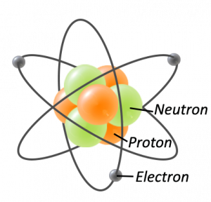 |
Nuclear = irradiation?
NO! Quite the contrary. In contrast to radiographies, mammographies, and scanners, MRIMRI means Magnetic Resonance Imaging but it can also mean the exam, the image, or the imager. Further information on MRI and its technique More (which is based on Nuclear Magnetic Resonance) does not use X-rays. There is no radioactivity. It is referred to as non-ionizing imaging. Is there any risk with MRIMRI means Magnetic Resonance Imaging but it can also mean the exam, the image, or the imager. Further information on MRI and its technique More? |
 |
What about MRIMRI means Magnetic Resonance Imaging but it can also mean the exam, the image, or the imager. Further information on MRI and its technique More? IRMLe terme IRM signifie Imagerie par Résonance Magnétique mais il peut désigner l'examen, l'image ou l'imageur.Pour en savoir + sur l'IRM et son principe More mainly focuses on the hydrogen nucleus 1 H, as it is strongly present in the human body in water molecules (H2O). He has only one proton and thus has the characteristic features cited above. |
|
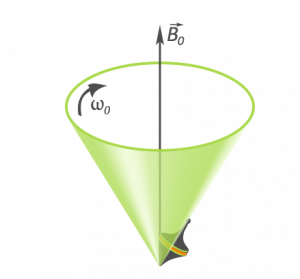
If we place those “magnetized” nuclei in a constant magnetic field B0, they start to rotate around the axis of the field like wooden tops, in a specified direction. We call it precession movement. The speed of the movement is set in the Larmor formula, which says that if B0 field increases, precession frequency, which is linked to speed, increases, and vice versa. |
 |
| The term “Magnetic Resonance” in NMR, is the result of another phenomenon in relation to the former described above. There can be an interaction between a radiofrequency wave (RF wave, i.e, magnetic field oscillating at a specified frequency), and the nuclei in precession: this is what we call resonance phenomenon. This resonance occurs if the RF wave has the same frequency as that of the precession of the spins. |
Do you know any other resonance phenomenons?
Yes! Who hasn’t heard of these two examples?
|
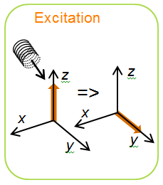
Through the resonance phenomenon, the RF wave provides energy to the spins: this is what we call excitation. With this added energy, spins start to orientate in another direction. This is magnetization shift. The angle of the magnetization shift is influenced by the intensity of the RF wave and the duration of the process.
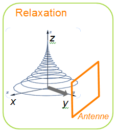
After the magnetization, spins have a natural tendency to return to their initial state, i.e, to the orientation they had in the B0 field axis. This is what we call relaxation. As spins rotate, their axis describes a sort of spiral (like a snail shell), so that they recover their equilibrium. At the same time, relaxation phenomenon radiates energy in the form of RF waves. It is during the relaxation phase that the NMR is recorded.
|
What about MRIMRI means Magnetic Resonance Imaging but it can also mean the exam, the image, or the imager. Further information on MRI and its technique More? In MRIMRI means Magnetic Resonance Imaging but it can also mean the exam, the image, or the imager. Further information on MRI and its technique More, this is exactly the same principle. We have the phases of:
|
|
|
|
|
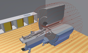

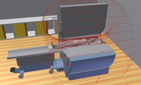
MRIMRI means Magnetic Resonance Imaging but it can also mean the exam, the image, or the imager. Further information on MRI and its technique More: the machine
Basically, a MRIMRI means Magnetic Resonance Imaging but it can also mean the exam, the image, or the imager. Further information on MRI and its technique More machine is:
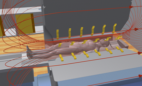
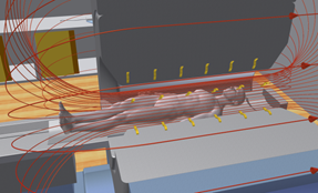
- A big magnet to generate a constant and uniform magnetic field B0
- Coils to create oscillating RF waves and to enable the excitation of the atoms.
- An antennaThe antenna is the device that emits radiofrequency impulses (for the creation of the signal) and/or receive the signal (for the image production). We mostly use a dedicated antenna according to the studied area (head antenna, knee antenna, etc.). More to measure the precession signal of the atoms that resonate. The antennaThe antenna is the device that emits radiofrequency impulses (for the creation of the signal) and/or receive the signal (for the image production). We mostly use a dedicated antenna according to the studied area (head antenna, knee antenna, etc.). More is positioned as close as possible to the area to be imaged.
The antennaThe antenna is the device that emits radiofrequency impulses (for the creation of the signal) and/or receive the signal (for the image production). We mostly use a dedicated antenna according to the studied area (head antenna, knee antenna, etc.). More is sometimes referred to as a transmitting receiving antennaThe antenna is the device that emits radiofrequency impulses (for the creation of the signal) and/or receive the signal (for the image production). We mostly use a dedicated antenna according to the studied area (head antenna, knee antenna, etc.). More. In that case, it generates the excitation RF waves and receives the relaxation RF waves. - A super PC that generates a computer algorithm that analyses all the measures.
Those elements are the minimum necessary to record the nuclear magnetic resonance signal.
In addition to that, three pairs of gradient coils are necessary, in the three directions of space, to make a slight variation in the magnetic field of the B0 field. This enables the machine to add a location data to the signal to give a position to the different signals, and thus create images (see chapter below).
Everything is then wrapped in shielding in order, as much as possible, to limit the magnetic field to small space.
Lastly, there can be a cooling system wif the MRIMRI means Magnetic Resonance Imaging but it can also mean the exam, the image, or the imager. Further information on MRI and its technique More is composed of superconducting magnets.
How are images generated?
|
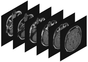
MRIMRI means Magnetic Resonance Imaging but it can also mean the exam, the image, or the imager. Further information on MRI and its technique More is an imaging technique that is able to generate visual “slices“ of human body in different planes. It is besides possible to obtain a reconstruction in three dimensions by assembling the slices. We saw above how to register the data from the NMN. Then, how are selected those data to generate an image? In order to know where the data comes from, it is necessary to make a spatial coding. This is the role of gradient coils. |
|
|
What is a gradient?
A field gradient is a slight linear variation over a given distance. When you add a field gradient to a magnetic field B0 you make the field vary in a given axis. In practice, if you take for example a field of 3 Tesla, the fact that you apply it with a field gradient could make it vary from 2,980 Tesla to 3,020 Tesla in a given direction. |
Several gradients are successively applied for the spatial coding, according to different axis (x, y, z). A first gradient, called “cut selection gradient” is applied. It enables it to determine a plane. Applying then a “phase gradient” enables it to move in the vertical axis of the plane. And then applying the “frequency gradient” enables it to move in the horizontal axis of the plane. |
 |
In order to generate the imaging, it is necessary to plan the temporal unfolding of the application of the different gradients and RF waves. The combination of gradients and RF waves are carefully organized in a sequence. The organization of the sequence will particularly enable it to obtain contrast, spatial resolution, and acquisition speed needed for the imaging. While the sequence is being applied, the NMR signal is recorded and stored “raw” in a sort of temporary image called K-space or Fourier plane. K-space is then mathematically treated through a Fourier transform in order to obtain an image of the studied area. |
MRIMRI means Magnetic Resonance Imaging but it can also mean the exam, the image, or the imager. Further information on MRI and its technique More: a technique with many elements to be studied and developed…
The explanations above give us a very short overview of the numerous parameters necessary to the production of MR images. There are still lots of other parameters.
Those parameters can influence the quality of the image, of the speed acquisition, of the exam duration. They can also modify contrasts in order to visualize organs or varied pathologies.
For some diagnosis it is necessary to inject a contrast agent. Then other elements must be taken into account.
It is also possible to make functional MRIMRI means Magnetic Resonance Imaging but it can also mean the exam, the image, or the imager. Further information on MRI and its technique More, or fIRM. It is an imaging method that enables one to visualize the brain activity “looking” at blood flows. We may then see which brain area is used for specific activities.
We can also make diffusion MRIMRI means Magnetic Resonance Imaging but it can also mean the exam, the image, or the imager. Further information on MRI and its technique More which focuses on the micro-movements of water molecules and enables one to study the organs structure to identify some pathologies.
Those are only few examples of what can be done with MRIMRI means Magnetic Resonance Imaging but it can also mean the exam, the image, or the imager. Further information on MRI and its technique More but it has still lots of other potentials.
All those parameters and applications of the MRIMRI means Magnetic Resonance Imaging but it can also mean the exam, the image, or the imager. Further information on MRI and its technique More make it a complex subject for which there are still many routes to investigate and many elements to improve.